Determination of elements concencetraions in human brain tissues using PIXE and EDX methods
Published in Journal of Radioanalytical and Nuclear Chemistry (2018) 318:2313–2319
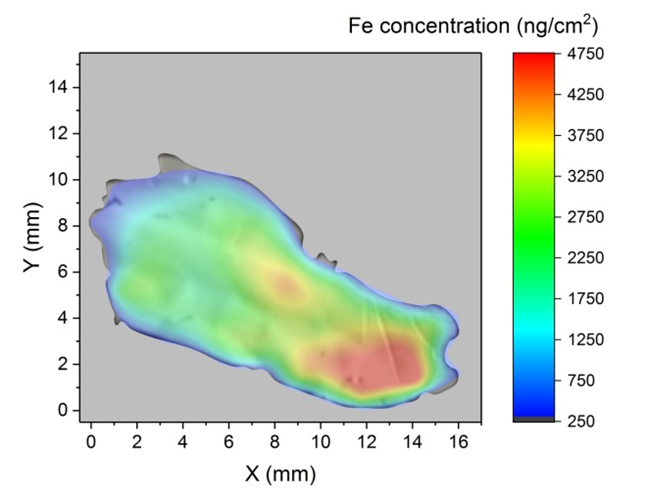
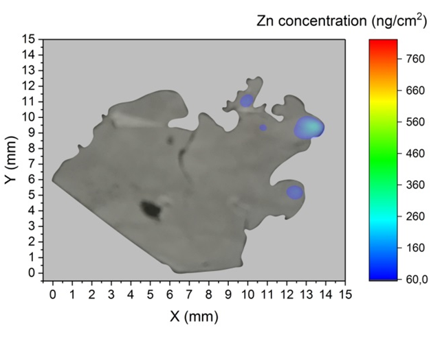
The most abundant heavy metal, iron, is stored in human body mainly in the brain tissue. Iron is a fundamental cofactor of many different enzymes in the human body and it is needed therefore for normal neuronal function. It is well known that metal ions in substantia nigra (SN) are associated with neuromelanin structure in substantia nigra pars compacta. Neurons store iron in the form of ferrous ions which is linked with neurodegenerative diseases such as Alzheimer’s and Parkinson’s disease, Friedreich’s ataxia, and Huntington’s disease. Post-mortem brain tissue sections from SN were obtained during the autopsy to prepare tissue sections for the pathology diagnosis at the Institute of Pathological Anatomy of the Comenius University. Original samples of substantia nigra pars compacta were first investigated by light microscopy and SEM (scanning electron microscopy) with EDX (Energy-Dispersive X-ray spectroscopy) for qualitative analyses. Four samples were analysed. Thin slices (5 μm) of the brain sectionswere placed on 2 μm Mylar foil and then fixed on frame holders. X-ray spectra from the PIXE analysis using 3 MeV proton beam (1 – 2 mm spot in diameter) with the intensity of ~ 3 nA with the sample tissue were collected. Up to 30 spots were measured on each sample. Fe, Mn, Cr and Zn concentration distribution maps were created. Maps with highest observed values for individual elements are shown in the figures (gray color represents the background level of the measurement).
SN samples with age and gender of donors | ||
|---|---|---|
| Sample | Age | Gender |
| S1 | 61 | Male |
| S2 | 76 | Male |
| S3 | 88 | Female |
| S4 | 90 | Male |